Bone Anatomy
Cortical vs. Trabecular Bone in Implant Osteotomy
A brief anatomy and biomechanics primer for implant clinicians, in Dr. Zvi Fudim's own words. Elasticity, rigidity, vascularization, and thermal behavior are as important as hardness - and shape every decision at the osteotomy stage.

Clinical primer · by Dr. Zvi Fudim
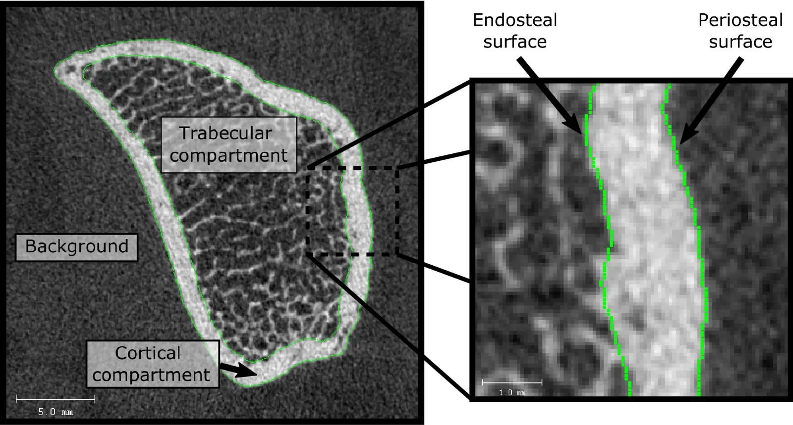
Cortical vs. trabecular bone.
It may seem like a trivial distinction, but the mechanical and biological differences between cortical and trabecular bone can surprise even experienced implant clinicians. Understanding these differences is fundamental to achieving precise osteotomies, minimizing trauma, and maximizing primary stability.
Elasticity
Elasticity is the ability of a material to return to its original shape after deformation. Trabecular bone can undergo elastic deformation of up to approximately 50%, whereas cortical bone typically deforms by only about 2% before reaching its limit.
This distinction has important clinical implications. Highly elastic materials require extremely sharp cutting instruments. Rather than being cleanly cut, elastic bone tends to deform around a dull drill, resulting in tearing instead of precise cutting. The consequence is an osteotomy with irregular walls, reduced dimensional accuracy, and a rougher internal surface. These imperfections may compromise the self-threading action of the implant and reduce primary stability.
Paradoxically, drilling elastic trabecular bone is not always easier. As the elastic walls tend to rebound toward the drill body, they generate increased lateral friction. Although trabecular bone appears softer, this sidewall friction can significantly increase heat generation if the cutting efficiency of the drill is inadequate.
Rigidity
Rigidity is essentially the opposite of elasticity and is a defining characteristic of cortical bone.
In many clinical situations, cortical bone is so rigid that a discrepancy of only a few microns can prevent an implant from reaching its fully seated position. Excessive compression of this rigid layer may also produce cortical microfractures, increasing surgical trauma and potentially prolonging the inflammatory phase of healing.
The combination of high hardness and rigidity often exceeds the practical cutting capacity of conventional stainless steel drills. As these drills lose sharpness through wear, friction increases, resulting in progressively greater heat generation and reduced cutting efficiency.
Vascularization
Perhaps the greatest biological difference between trabecular and cortical bone lies in their vascular architecture.
Trabecular bone is highly vascularized and contains abundant blood vessels, marrow, plasma, and cellular elements that play a critical role in healing and bone regeneration. Excessive irrigation during osteotomy preparation may wash away these biological components and can also reduce the natural lubrication produced by blood, particularly during osseodensification procedures where frictional interaction with bone is intentional.
Cortical bone is fundamentally different. Its blood supply is relatively limited and is derived primarily from the periosteum and intracortical vascular channels. The periosteum and cortical bone are also richly innervated with sensory nerve fibers, making cortical trauma one of the principal sources of postoperative pain during implant surgery.
Summary
Many practitioners consider hardness to be the only meaningful difference between cortical and trabecular bone. In reality, these tissues differ in elasticity, rigidity, vascularization, thermal behavior, and biological response to surgical trauma. As a result, they should not be approached as though they are the same material.
The Crown Down implant drill kit was developed around this principle. Rather than preparing both bone types with a single drilling strategy, the invention recognizes that each tissue has unique mechanical and biological characteristics. By first creating precise clearance through the rigid cortical layer with dedicated tungsten carbide cortical drills, and then preparing the elastic trabecular bone with a separate undersized drill, each tissue is treated according to its own properties.
This tissue-specific approach reduces friction where heat is most likely to develop, preserves the structural integrity of the trabecular bone, minimizes cortical trauma, improves osteotomy precision, and helps achieve predictable primary implant stability while maintaining the biological conditions necessary for optimal healing.
Why is hardness alone the wrong way to compare cortical and trabecular bone?
The clinical shorthand for “bone quality” is Carl Misch’s D1–D4 classification, which maps neatly onto the ratio of cortical to trabecular bone at the implant site. D1 (dense cortical throughout, typical of the anterior mandible) is almost all cortical; D4 (a thin cortical shell over fine low-density trabecular, typical of the posterior maxilla) is dominated by the elastic, poorly-supportive interior. The two extremes ask for opposite things from a drill sequence, and yet most conventional protocols use the same pilot-to-final ascending drills for both.
This is where the elasticity data above becomes practical. The ~50% elastic reserve in trabecular bone means the wall rebounds onto the drill as soon as the flute passes. A dull edge doesn’t cut that rebounded wall - it burnishes it, produces heat by lateral friction, and leaves an osteotomy with irregular internal geometry. The clinician interprets the softer bone as “easier” and often under-irrigates, which compounds the heat problem. The higher-leverage variable is not saline volume, it’s the cutting edge itself.
Conversely, the ~2% elastic limit of cortical bone means it does not rebound - it fractures. Excessive compression during the pilot-to-final ascending sequence, especially with an under-sharp final drill, is what produces the cortical microfracturing that shows up on histology after implant failure. The remedy is not more torque; it is a drill that clears the cortex cleanly in one pass at the correct diameter.
Vascularization: the biology most drilling protocols quietly wash away.
Trabecular bone is not just soft - it is where the biology of osseointegration lives. Bone marrow spaces contain osteoblast precursors, mesenchymal stem cells, vascular endothelial cells, plasma, and the paracrine signaling that drives early peri-implant bone formation. The osteotomy wall is not a passive container for the implant; it is the active biological interface across which integration happens.
Continuous copious saline irrigation is the standard response to the heat threshold established by Eriksson and Albrektsson in 1983 (47 °C for 60 seconds), and it does its job for heat - but the same saline stream that carries heat away also dilutes and displaces the biology of the trabecular wall. This is a trade the conventional protocol accepts because it has no alternative: stainless steel drills generate the friction they generate, and irrigation is the only lever available.
A drill material with materially higher thermal conductivity removes the requirement. Solid tungsten carbide conducts heat at about 110 W/m·K, roughly six times steel; combined with the 200 RPM clinical protocol speed and a 2-drill protocol that reduces the number of passes through the trabecular wall to one, the heat budget stays inside the ~41°C presumed mouth temperature envelope Crown Down measured on the bench, comfortably below the 47°C thermal-injury reference, without saline having to displace the biology.
Autogenous bone chips collected on the flutes of a dry-drilled Crown Down cortical drill are a downstream benefit of the same architecture: undiluted, biologically viable, and available for immediate re-implantation at the same surgical site.
How does bone innervation affect postoperative implant pain?
Trabecular bone is essentially aneural. The periosteum overlying cortical bone and the cortex itself carry a dense sensory nerve supply, which is why every clinician working without adequate anesthesia has heard the same complaint: cortical prep hurts and trabecular prep does not. The clinical takeaway is not just about intra-operative analgesia; it is that postoperative pain, inflammation, and prostaglandin release track cortical trauma more than trabecular trauma.
Minimizing cortical passes - fewer drills through the cortex, at lower RPM, generating less heat - is directly correlated with a smoother postoperative course. The Crown Down protocol’s single cortical drill, matched to the final implant diameter and used at approximately 250 RPM, is designed around exactly this objective. The cortex is opened once, not five to eight times, and the tissue that carries the sensory nerve supply is exposed to the minimum practical mechanical and thermal insult.
Why do cortical and trabecular bone need different drilling instruments?
There is a general principle in surgery, borrowed from engineering: an instrument optimized for two different substrates is optimized for neither. The conventional pilot-to-final ascending drilling sequence treats cortical and trabecular bone as though they are one continuous tissue with a hardness gradient. They are not. They are two different tissues with different elastic behavior, different vascular architecture, different innervation, and different biological roles in osseointegration.
The Crown Down 2-drill kit is a direct application of that principle. The cortical drill (drill #1) is matched to the implant diameter, made of solid tungsten carbide, and used first to open the rigid crestal layer in a single clean pass without countersinking. The trabecular drill (drill #2) is smaller than the implant diameter, tuned for the elastic wall behavior of the interior, and used to shape the site while preserving the biological components that drive integration. Two tissues, two drills, two protocols. The full range of drill sizes is on the dental implant drills page.
The full 2-drill protocol, including RPM, torque, and irrigation guidance for both drills, is documented separately. The comparison to osseodensification and osseocompression covers where controlled bone compaction fits into the trabecular-preparation half of this argument.
Frequently asked questions
Quick answers to questions clinicians ask most about this topic.
Keep reading
Related reading
Explore related pages on the Crown Down dental implant drilling kit, protocol, and clinical science.
Protocol
The 2-Drill Implant Osteotomy Protocol
How drill #1 and drill #2 prepare the implant osteotomy in two steps.
Read moreCortical Drill
Cortical Drill for Dental Implants
Drill #1 in the Crown Down 2-drill protocol: matched per implant diameter, used first to clear dense crestal bone.
Read morePrimary Stability
Osseodensification vs. Osseocompression
Bone-chip compaction vs. trabecular compression for implant primary stability.
Read more